
Figure 2.
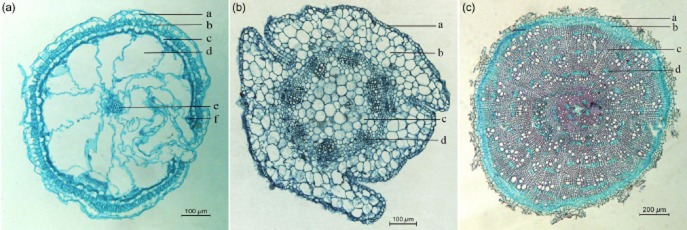
Transverse sections of leaf, stem and root of Halogeton glomeratus seedlings. (a) Leaf, exhibiting epidermal cells (a), palisade tissue (b), lignified cells (c), water-storage tissue (d), vascular bundle (e) and small peripheral bundles (f). (b) Stem, showing epidermal cells (a), cortex (b), hollow pith (c) and collateral vascular bundle (d). (c) Root, exhibiting cortex (a), secondary phloem (b), xylem (c) and large parenchyma cells (d).