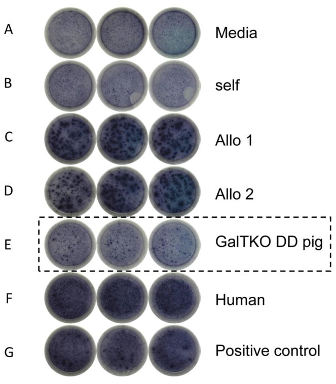
Figure 4. ELISPOT assay for INFγ.
Many spots could be seen in the wells stimulated by allogeneic cells (C and D), human cells (F) and PHA (G) on POD183 (B336), while the spots were few in the well stimulated by GalTKO DD pig cells (E). This was similar to what was observed in wells stimulated by media (A) and autologous PBMC (B).

