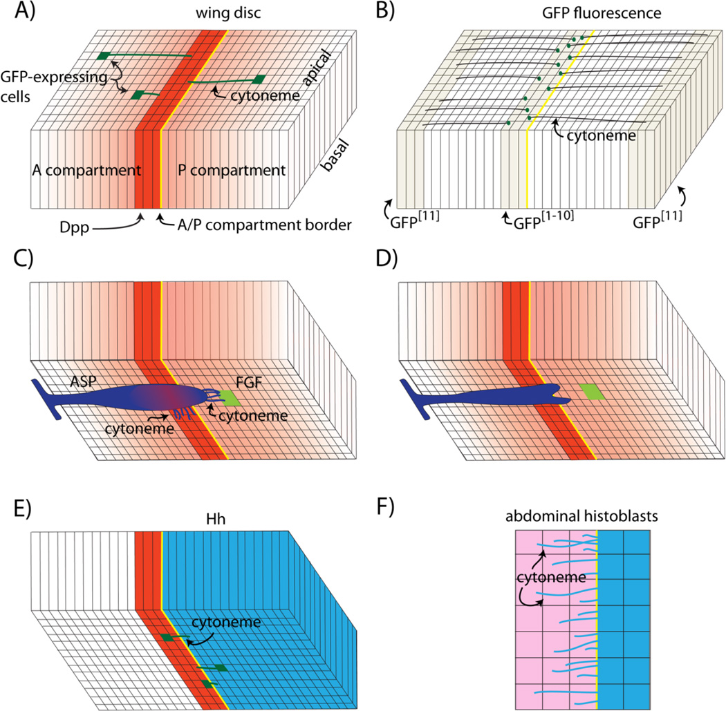
Figure 1.
Cytonemes of the Drosophila wing disc and histoblasts. A: The wing blade primordium is depicted as a columnar monolayer subdivided by the anterior/posterior compartment border (yellow) and a strip of Dpp-expressing cells (red). Concentration gradients of Dpp protein and Dpp signal transduction (red) spread from the Dpp expression domain and cell clones that express GFP have cytonemes (green) that orient along the apical surface toward the Dpp expressing cells. B: GFP reconstitution (GRASP) generates fluorescence (green dots) in the Dpp expression domain in wing discs that express complementary parts of GFP as membrane-tethered external proteins in the disc flanks and the Dpp expression domain. Fluorescence marks points of cell-cytoneme contact. C: The tracheal air sac primordium (ASP; purple) adjoins the basal surface of the wing disc and extends cytonemes toward both Dpp-expressing (red) and FGF-expressing (green) cells; the gradient of Dpp signal transduction in the ASP is shown in red. D: Mutant ASP cells that do not extend cytonemes that synapse with the disc cells do not activate Dpp signal transduction and the ASP is morphologically abnormal. E: Basal cytonemes (green) that carry Hh extend from both Hh-expressing cells (blue) in the posterior compartment and from Hh-receiving cells (red) in the anterior compartment. F: The rows of Hh-expressing cells (blue) in monolayered epithelium of abdominal histoblasts extend Hh-carrying cytonemes to Hh-receiving cells (pink) that express the Hh receptor Patched (Ptc).

