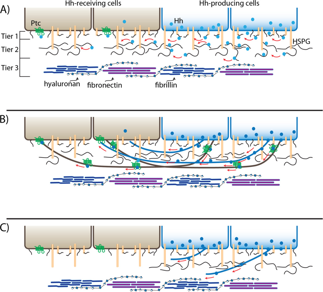
Figure 2.
Restricted diffusion and cytoneme models of Hh gradient formation. A: Drawings depicting two cells expressing and secreting Hh (blue) that interacts directly with HSPGs in the ECM and disperses to generate a concentration gradient across two receiving cells (brown; Ptc, green) by surface diffusion. The "conceptual” ECM is depicted as three tiers consisting of (1) glycolipids (black lines) and surface glycoproteins (tan cylinders with black lines), (2) HSPGs (tall tan cylinders with black lines) and (3) extracellular microfibils (blue), fibronectin (purple) and hyaluronan (beaded strand). B: This drawing depicts Hh dispersing along cytonemes (black) that extend from expressing (brown) and receiving (blue) cells. C: Mutant receiving cells (brown) that do not express HSPGs neither extend cytonemes nor are contacted by cytonemes that extend from Hh-expressing cells.

