
Fig 6. Key residues identified in DnaK SBD.
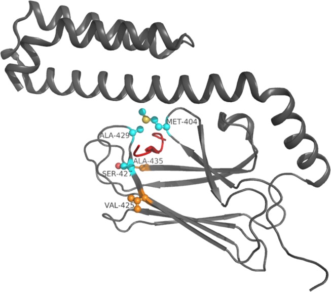
Residue side-chains are in ball and stick, Cα atoms of residues previously exploited by known inhibitors are in light blue, Cα of the new identified hot spot residues are in orange. The Api88 peptide is represented in red.