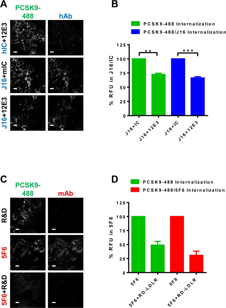
Fig 3. Anti-APLP2 or Anti-LDLR antibody effects on PCSK9 internalization.
(A) PCSK9-488 internalization in the presence of 12E3 and hIC (top row), mIC and J16 (middle row) or 12E3 and J16 (bottom row). Internalized human antibodies shown in blue. Scale bars, 10 μM. (B) Quantification of (A) shown as average percent signal of PCSK9-488 per cell of PCSK9-488/J16 (green bars) or average percent signal of J16 per cell of PCSK9-488/J16 (blue bars) with SEM from 3 independent experiments. (C) PCSK9-488 internalization in the presence of RD-LDLR (top row), hIC and 5F6 (middle row) or RD-LDLR and 5F6 (bottom row). Internalized mouse antibodies shown red. Scale bars, 10 μM. (D) Quantification of (C) shown as average percent signal of PCSK9-488 per cell of PCSK9-488/5F6 (green bars) or average percent signal of 5F6 per cell of PCSK9-488/5F6 (red bars) with SEM from 3 independent experiments.

