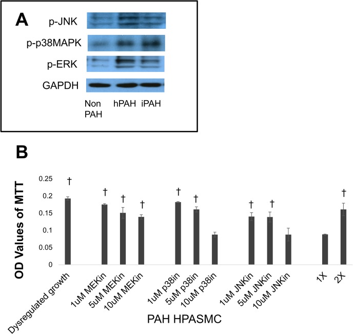
Fig 4. PAH HPASMC proliferation via MAP kinases.
(A) Western blot showing levels of phospho-JNK, phospho-p38MAPK, phospho-ERK and GAPDH after overnight incubation of the cells in medium containing 0.2% FBS. (B) Twenty four hours after seeding, the growth medium was replaced with quiescence medium and inhibitors (MEKin = U0126; p38in = SB203580; JNKin = SP600125) at respective concentrations were added to the culture. Cell proliferation was determined 5 days later using the MTT assay. Bar graphs represent the average OD values from triplicate wells. The error bars represent standard deviation. This figure shows results from an hPAH cell strain and is representative of results obtained from other PAH cell strains. †p < 0.001 vs 1X cell number.

