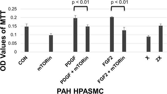
Fig 8. PAH HPASMC dysregulated and growth factor stimulated proliferation.

Twenty four hours after seeding, medium was replaced with quiescence medium. Cells were preincubated for 30 min with 10 ng/ml rapamycin (mTORin) and then treated with or without 10 ng/ml PDGF or FGF2. Cell proliferation was determined 5 days later using the MTT assay. Bar graphs represent the average OD values from triplicate wells. The error bars are the standard deviation. This figure is from an hPAH cell strain and is representative of results obtained from other PAH cell strains.
