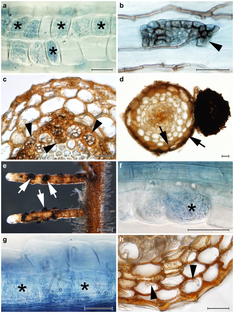
Fig 1. The colonization patterns observed in Norway spruce (Picea abies) and European blueberry (Vaccinium myrtillus) roots in Experiment 1.
1a) Typical ericoid mycorrhizal colonization formed by Rhizoscyphus ericae in blueberry roots (asterisks); stained with trypan blue, observed with DIC, bar = 25 μm. 1b) An intracellular microsclerotium formed by Phialocephala helvetica in a blueberry root (arrowhead); stained with trypan blue, observed with DIC, bar = 25 μm. 1c) Intracellular microsclerotia formed by P. helvetica in the vascular cylinder of a spruce root (arrowheads); observed with DIC, bar = 25 μm. 1d) A Hartig net formed within the spruce root cortex (arrows) and an extraradical sclerotium formed on the spruce root surface (asterisk) by Acephala macrosclerotiorum; observed with DIC, bar = 25 μm. 1e) Spruce root tips colonized by A. macrosclerotiorum with extraradical superficial sclerotia formed on the root surface (arrows); bar = 0.5 mm. 1f) Intracellular hyphal loops morphologically resembling ericoid mycorrhizae (asterisks) formed by A. macrosclerotiorum in blueberry roots; stained with trypan blue, observed with DIC, bar = 25 μm. 1g) Loose intracellular hyphal loops which may morphologically resemble ericoid mycorrhiza (asterisks) formed by Phialocephala glacialis in blueberry roots; stained with trypan blue, bar = 25 μm. 1h) Intracellular colonization of spruce root cortex by P. glacialis (arrowheads); bar = 25 μm.

