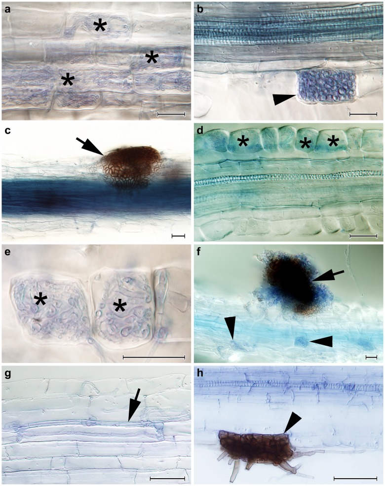
Fig 2. The colonization patterns observed in European blueberry (Vaccinium myrtillus) roots in Experiment 2 and in silver birch in Experiment 3.
2a) Intracellular hyphal colonization resembling ericoid mycorrhiza formed by Acephala applanta AAP-1 in blueberry roots (asterisks). 2b) An early stage of the development of an intracellular microsclerotium formed by A. applanata AAP-1 in a blueberry root (arrowhead). 2c) An extraradical sclerotium formed on the surface of a blueberry root by A. applanata AAP-1 (arrow). 2d) A blueberry hair root colonized in a manner resembling ericoid mycorrhiza by Acephala macrosclerotiorum AMA-11 (asterisks). 2e) A detail of two blueberry rhizodermal cells intracellularly colonized by A. macrosclerotiorum AMA-11 in a manner resembling ericoid mycorrhiza (asterisks). 2f) An extraradical sclerotium formed on the surface of a blueberry root by A. macrosclerotiorum AMA-11 (arrow). Note accompanying intracellular hyphal colonization (arrowheads). 2g) A loose intracellular hyphal loop formed by A. macrosclerotiorum AMA-1 in a birch root (arrow). 2h) A melanised intracellular microsclerotium formed by Acephala applanata AAP-1 in birch (arrowhead). All figures stained with trypan blue, observed with DIC, bars = 25 μm.

