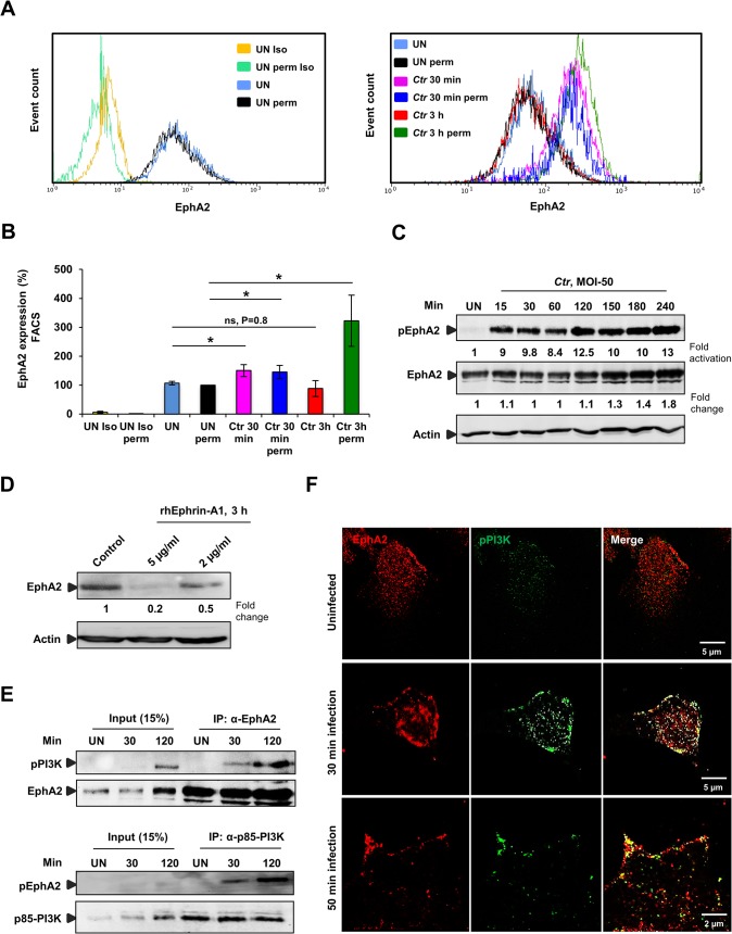
Fig 3. Ctr-induces EphA2 activation and receptor internalization which associates with pPI3K during early infection.
(A) HeLa cells uninfected (UN) or infected with Ctr (MOI-50-75) for 30 min or 3 h were analyzed via FACS for surface EphA2 under non permeabilised or total EphA2 under permeabilised condition. Controls were indicated on the left curve chart and corresponding samples on the right curve chart of the same experiment. (UN: uninfected, Iso: isotype and perm: permeabilised). (B) Result of experiment shown in A demonstrated as bar chart. Shown is the mean ± SD of three independent experiments normalized to UN perm. *P<0.05, ns: non-significant. Error bars show mean ± SD. (C) HeLa cells were UN or infected with Ctr (MOI-50) for the indicated time points. The cells were immunobloted against pEphA2 and Actin. The blot was stripped and reprobed for total EphA2. (D) HeLa cells were treated with control or rhEphrin-A1 for 3 h and were harvested for WB analysis. (E) HeLa cells were UN or infected with EB for the indicated time points and immunoprecipitated (IP) with α-EphA2 or α-p85-PI3K antibodies. The IP material was solved in 40 μl Laemmli (100%) and loaded 20 μl for WB studies (50%) or (F) immunostained using α-EphA2 and α-pPI3K for 2 h at RT followed by secondary staining with anti-mouse Alexa fluor 488 and anti-rabbit Alexa fluor 647 for the microscopic analysis, respectively. Magnification is indicated in size bar.

