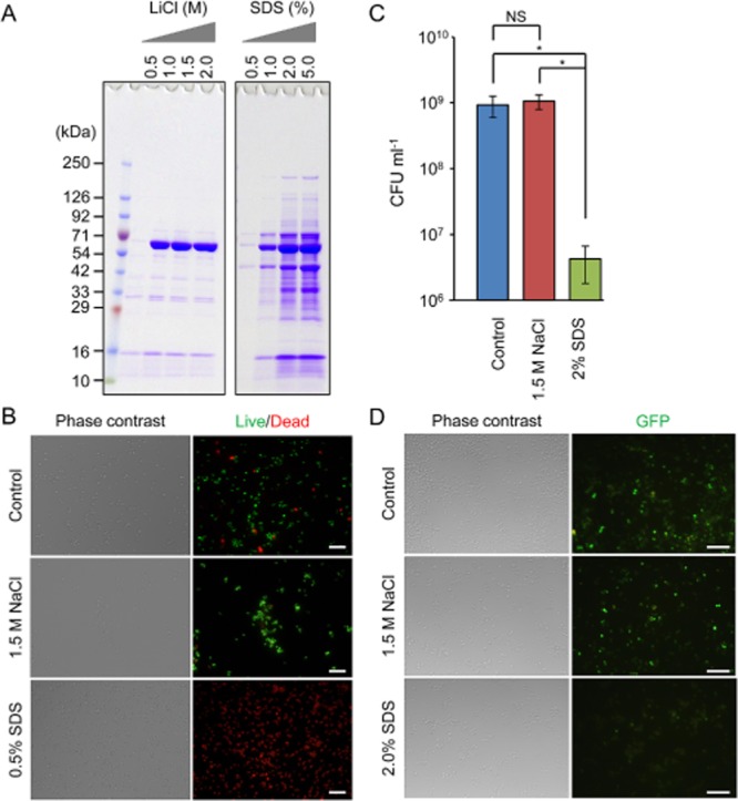
Fig 2.

Assessment of cytotoxicity of NaCl and SDS.A. Extracellular matrices of S. aureus MR23 were extracted by the addition of the indicated concentrations of LiCl or SDS and were applied to SDS-PAGE with CBB staining. A molecular mass marker was also loaded to the left lane.B. Live/Dead staining images of MR23 biofilm cells after the treatment with 1.5 M NaCl or 0.5% (w/v) SDS are shown. A solution of 0.9% NaCl was used as a control. Phase contrast and fluorescence images are shown. Live and dead cells are stained in green and red respectively.C. Colony-forming unit of biofilm cells before and after the treatment with 1.5 M NaCl or 2% (w/v) SDS were measured. The means and standard deviations of triplicate determinations are represented.D. Staphylococcus aureus MR23 pP1GFP cells were treated with 1.5 M NaCl, 2% (w/v) SDS or PBS (as control) was observed with a fluorescence and phase-contrast microscope. Bars indicate 10 μm (B and D).
