Fig 4.

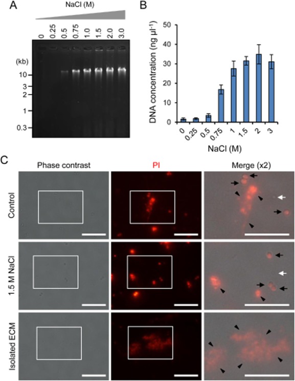
Extracellular DNAs are detached from biofilms by the addition of NaCl and harvested quantitatively.A. Extracellular matrices from S. aureus MS10 biofilms were extracted with the indicated concentrations of NaCl and were subjected to agarose gel electrophoresis. The gel was stained with ethidium bromide. The positions of molecular mass markers in kilobase pairs (kb) are shown at the left.B. Extracellular DNAs in the extracted ECM were purified to remove contaminated proteins and quantified by measuring absorbance at 260 nm. The means and standard deviations of triplicate determinations are represented.C. MS10 biofilm cells treated with or without 1.5 M NaCl and the extracted ECM were stained with PI and were observed with a fluorescence and phase-contrast microscope. Higher magnification (twofold) images of the white rectangles in the phase contrast and fluorescence images are merged. Black arrows, white arrows and black arrowheads represent bacterial cells stained with PI (dead cells), non-stained cells (variable cells) and eDNAs respectively. Bars indicate 10 μm.
