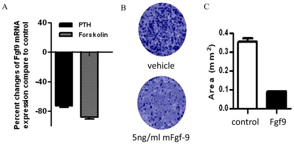
Figure 4.

(A) Effects of PTH and forskolin treatment on Fgf9 expression in primary BMSC cultures. Data were normalized to GAPDH RNA expression in the same sample and expressed as percent changes compared to controls. (B) Effect of continuous treatment of BMSCs with murine Fgf9 assessed by VK staining. VK-positive mineralized nodules were stained black. (C) Exposure of Fgf9 from Day 0 to day 20 increased VK staining area. Total colony area positive for VK staining was quantified as area (mm2). The results of these assays were confirmed by repeating the experiment three times. All data are shown as mean ± SD. *: p<0.05.
