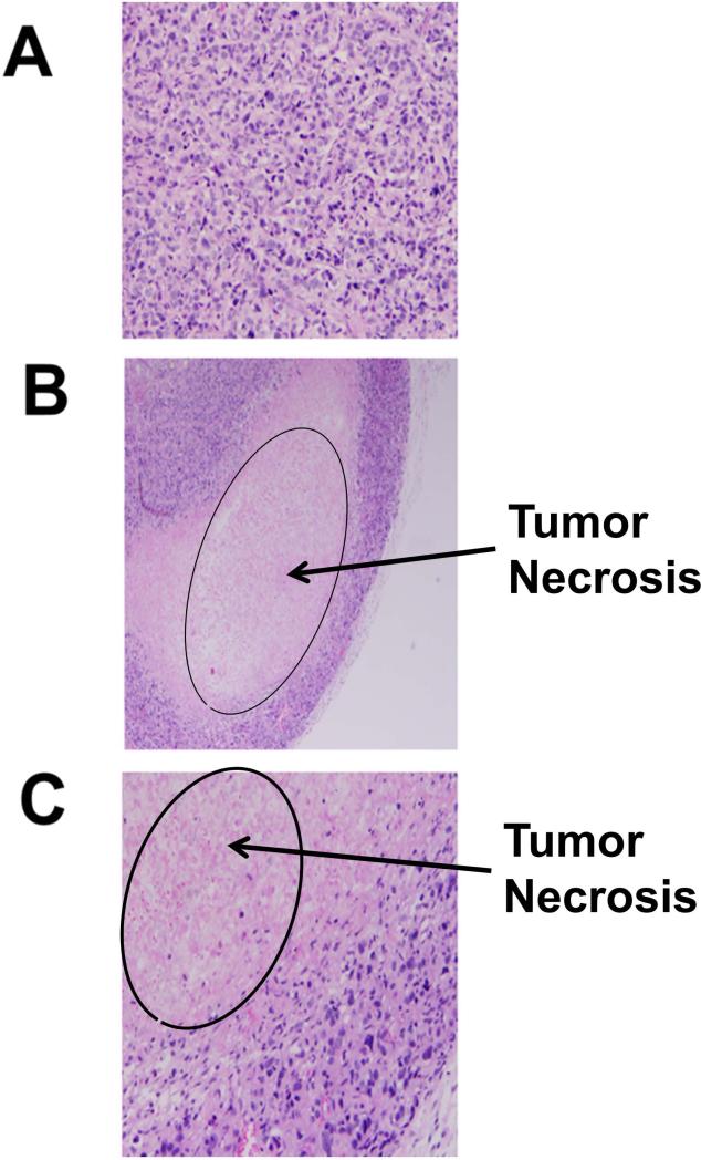
Figure 6.
H and E staining of tumor sections of mouse with and without compound treatments. A) Tumor tissue without any compound treatment, B) compound 5 treatment, C) compound 9 treatment. Sections from treated tumors (B, C) showed areas of necrosis that presented as eosinophilic debris with no cellular structures and nuclei.

