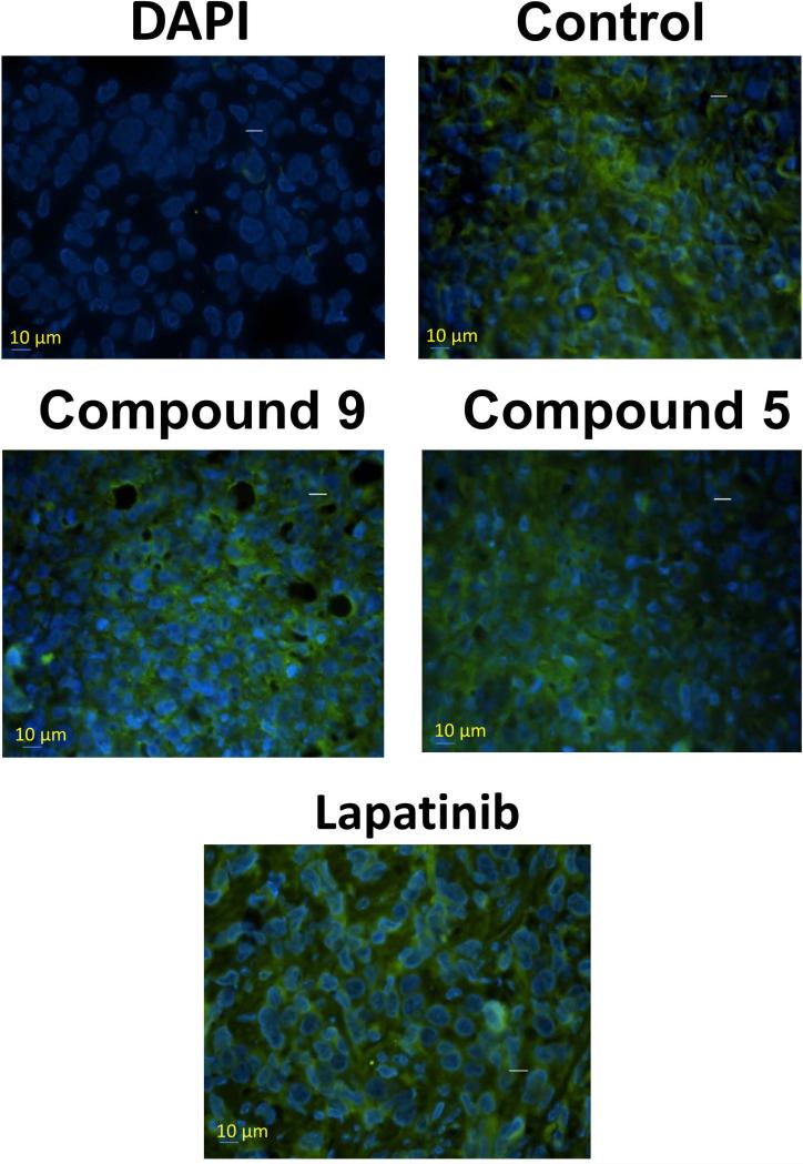
Figure 9.
HER2 expression in tumor sections of mice evaluated by fluorescently labeled anti-HER2 antibody (green fluorescence). Tissue section without any antibody treatment and labeled with nuclear stain DAPI. Tissue section without any compound treatment, compounds 9, 5, and lapatinib treatment are shown. Notice the HER2 expression in all the tumors suggesting that compounds do not affect the expression of HER2 protein.

