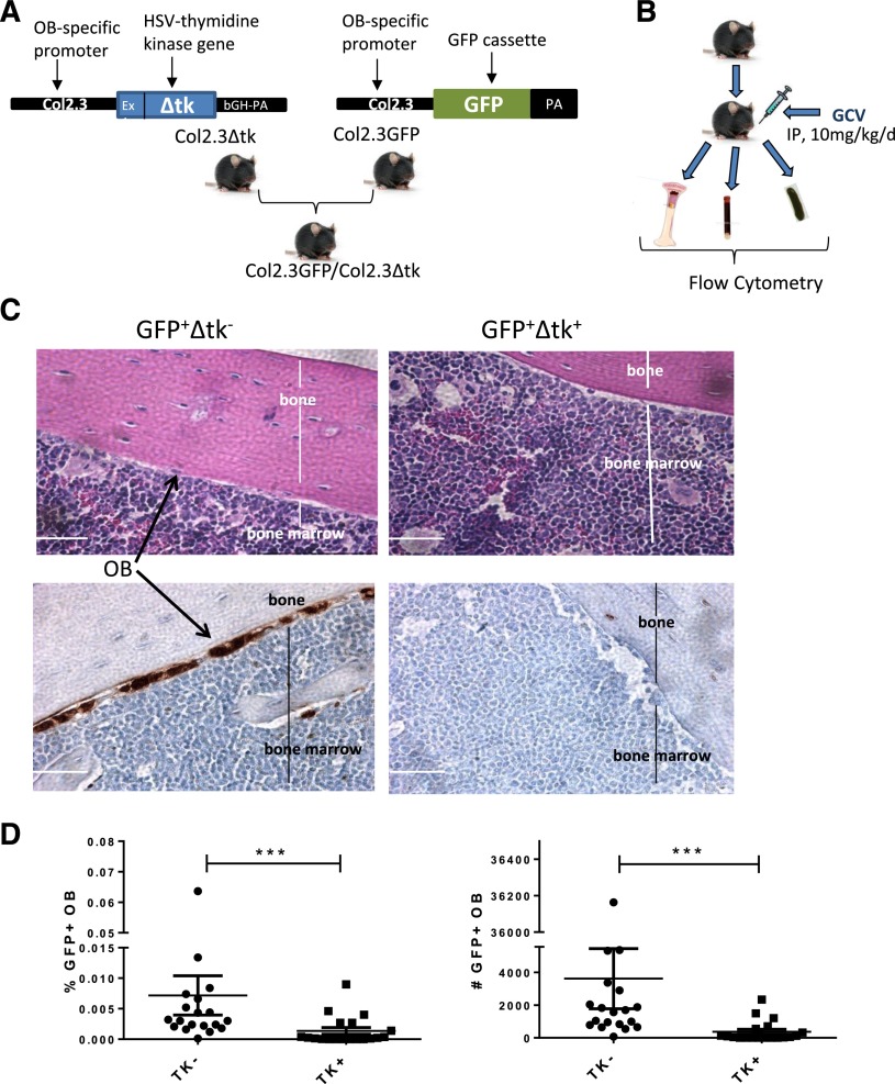
Figure 1.
Ablation of BM OBs in Col2.3GFP/Col2.3Δtk mice. (A) Strategy for generation of GFP-expressing OB ablation mice. (B) Experimental procedure for OB ablation and detection. (C) (Upper) Hematoxylin and eosin staining and (lower) GFP and DAPI staining of femur sections demonstrating the presence and absence of OB following GCV treatment in control (TK−) and ablation (TK+) mice, respectively. (D) Percentage (left) and number (right) of OBs in BM following GCV treatment as detected by flow cytometry (n = 19). Error bars represent mean ± SEM. Significance values: ***P < .001.

