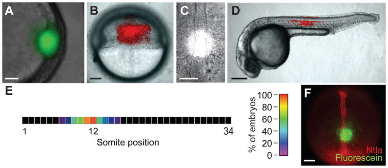
Figure 7. Localized irradiation of zebrafish embryos.

(A) Embryos injected with 2 nL of a 0.05% cFD solution were irradiated within the shield at 6 hpf for 10 sec using a circular photomask (100-μm diameter). As expected, a circular region of green fluorescence was immediately apparent in the targeted region. (B) Embryos injected with 50 pg of Kaede mRNA and irradiated laterally at 6 hpf for 10 sec using a rectangular photomask (200 × 300 μm). A rectangular region of red fluorescence was immediately observed in the targeted region. (C) Brightfield micrograph of a 10-hpf embryo undergoing UV irradiation through a circular, 100-μm-diameter photomask positioned 100 μm above the posterior end of the chordamesoderm. Grid overlays using Metamorph® software are not shown. (D) Embryos injected with 50 pg of Kaede mRNA and locally irradiated as in (C). A red fluorescent region of notochord and floor plate cells centered around the twelfth somite was visible at 1 dpf. (E) Heat map demonstrating the precision with which zebrafish embryos can be locally irradiated as described in (C). The average location of red fluorescent notochord cells along the anterior-posterior axis resulting from the targeted irradiation of 10-hpf embryos is shown (n = 18 embryos). (F) Fluorescence micrograph of a 10-hpf embryo injected with cFD, irradiate as described in (C), immediately fixed with paraformaldehyde, and immunostained with anti-Ntla and anti-fluorescein antibodies. A circular region of uncaged fluorescein was detected within the Ntla-expressing chordamesoderm, 100 μm anterior to the tailbud. Scale bars: A and C, 50 μm; B, 100 μm; D, 200 μm; F, 100 μm.
