
Fig. 6.
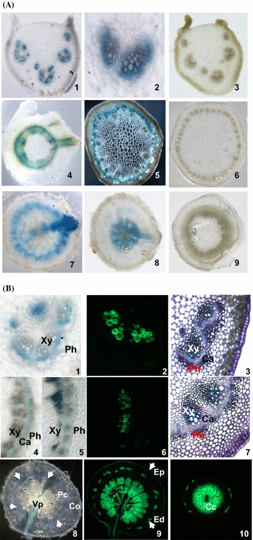
Localisation of Bn-FAE1.1 expression in vascular tissues of B. napus. a GUS activity was visualised in hand-cut transverse sections of roots, stems and leaf petioles of young B. napus plants transformed with the Bn-pFAE1.1 837:GUS construct. 1–3 Leaf petioles showing staining restricted to vascular bundles. 4–6 Stems. 4 Upper stem with emergent leaf vasculature. 5 Mid stem, showing fasicular staining in dark field. 7–9 Roots. 7 Mid root with emerging lateral root. 8 Lower root. 3, 6, 9 Stained sections from a non-transformed control plant. b Vibratome-cut transverse sections (100 µ) of leaf petioles (1–3) 1 GUS staining in zone between xylem and phloem; mid stem (4–7); mid root (8, 9), lower root (10). Transverse sections 3 and 7 are stained with toluidine blue. Sections 2, 6, 9 and 10 are stained with berberine hemi-sulphate to visualise suberin aromatic component, counter-stained with aniline and visualised under UV light. Transverse sections 1, 4, 5 and 8 are stained for GUS activity. White arrows indicate extent of annulus of GUS staining in 8. Co cortex, Ed endodermis, Ep epidermis, Pc pericycle, Cc central cycle, Vp vascular pole, Xy Xylem, Ca vascular cambium, Ph phloem