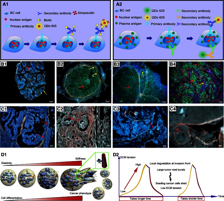
Fig. 2.
QDs-based imaging for studying BC biomarker interactions. Schematic plots of QDs-based single (A1) and double biomarkers imaging (A2). Decrease of IV collagen (yellow arrows) with the increase of HER2 (red arrows): HER2 (0), intact IV collagen (B1); HER2 (+), IV collagen becomes unsmooth and thinner (B2); HER2 (2 +), IV collagen becomes degraded (B3); HER2 (3+), complete IV collagen degradation (B4). BC invasion patterns: Washing Pattern (C1), Ameba-like Pattern (C2), Polarity Pattern (C3), and Linear Pattern (C4). Dynamic changes of IV collagen implied the pulse-mode of cancer invasion and metastasis: expansive growth of tumor nests burst to form disseminations (D1), the process from growth to burst is more quickly than the last (D2). ECM: extracellular matrix. Reproduced with permission from Ref [31–33]

