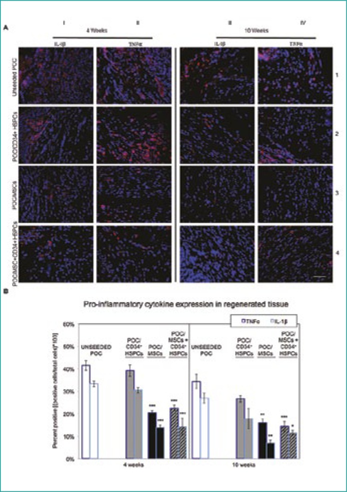
Figure 3.
Pro-inflammatory IL-1β and TNFα cytokine expression in regenerating bladder tissue.
(A) Detrimental to proper tissue regeneration is the presence of confounding pro-inflammatory cytokines including IL-1β and TNFα. IL-1β (red) and TNFα (red) levels were highly abundant in CD34+ HSPC and uPOC grafts at the 4 week time-point. TNFα levels remained elevated 10 weeks post-surgery while IL-1β decreased only moderately in the unseeded samples. IL-1β and TNFα levels in POC/MSC and POC/MSC + CD34+ HSPC grafts were approximately half the value found in POC/CD34+ HSPC and uPOC grafts at the 4 week time-point. There was a general decrease in pro-inflammatory cytokine expression at the 10 week time-point for all samples; however, TNFα expression was relatively elevated with the POC/CD34+ HSPC seeded construct. n = 10 images/animal. Blue = DAPI. Magnification, 400x. Scale bar, 50 µm.
(B) Quantification of pro-inflammatory cytokine expression, shown as percentage of cells staining positive for TNFα (left bars) and IL-1β (right bars) at 4 weeks (left panel) and 10 weeks (right panel). The cytokine expression profile for uPOC graft tissue was predominantly pro-inflammatory, with mean percentages of 41.5 ±2.2% TNFα+ and 33.3 ±1.2% IL-1β+ at 4 weeks and 34.3 ±3.1% TNFα+ and 27.0 ±2.3% IL-1β+ at 10 weeks. Significant reduction in both TNFα and IL-1β expression was observed for grafts seeded with MSCs or MSCs/CD34+ HSPCs (4W: POC/MSC 20.5 ±0.8% TNFα+ and 13.9 ±0.9% IL-1β+, POC/MSC + CD34+ HSPCs 22.5 ±1.3% TNFα+ and 14.2 ±3.7% IL-1β+; 10W: POC/MSC 16.0 ±1.6% TNFα+ and 7.0 ±1.2% IL-1β+, POC/MSC + CD34+ HSPCs 14.4 ±2.2% TNFα+ and 11.5 ±1.1% IL-1β+); grafts seeded only with CD34+ HSPCs did not show this effect (4W: 39.2 ±2.6% TNFα+ and 30.5 ±1.3% IL-1β+; 10W: 26.6 ±1.5% TNFα+ and 17.8 ±4.6% IL-1β+). Data shown as means ±SE. Significance shown for comparison of cell-seeded groups to uPOC; *P <0.05, **P <0.01, ***P ≤0.001, ****P <0.0001.

