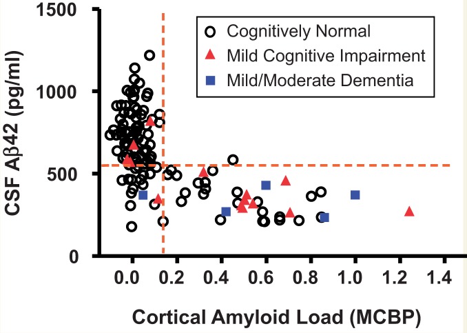
Figure 1.
Illustration of the association between concentrations of CSF amyloid-β42 and cortical amyloid load as revealed by PET. Research participants in the Knight Alzheimer’s Disease Research Centre underwent clinical assessment, CSF collection by lumbar puncture and amyloid imaging by PIB PET within a 12-month period. Cortical amyloid load is presented as the mean cortical binding potential (MCBP) calculated from the prefrontal cortex, precuneus, lateral temporal cortex and gyrus rectus, with cerebellum (very low PIB binding) as the reference region. CSF amyloid-β42 (Aβ42) values were obtained with the INNOTEST® ELISA kit (Fujirebio, formerly Innogenetics). Dashed lines illustrate potential cut-offs for PIB (right of the vertical line) and CSF amyloid-β42 (below the horizontal line) positivity. The cohort (age ≥ 65 years) included 113 cognitively normal participants, 14 with mild cognitive impairment/very mild dementia, and five with mild/moderate dementia. The majority of PIB+ individuals had low CSF Aβ42 whereas the majority of PIB− individuals had high levels of CSF Aβ42. All but one of the ‘discordant’ values are to be found in the lower left quadrant (low CSF Aβ42/low PIB). Concordance is observed in symptomatic and asymptomatic (presumed pre-symptomatic) individuals. Symptomatic amyloid-negative individuals may have non-Alzheimer aetiologies. Reprinted from Advances in Medical Sciences, Biomarkers of Alzheimer's disease and mild cognitive impairment: A current perspective, available online 9 December 2014, with permission from Elsevier.

