. 2015 Apr 2;4:e06828. doi: 10.7554/eLife.06828

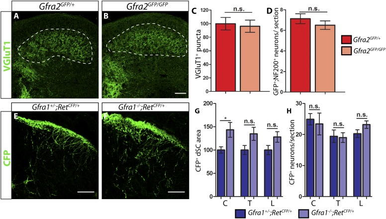
Figure 1. P7 Gfra2 mutant mice show normal dorsal spinal cord (dSC) VGLUT1 staining and Gfra1 null mice display normal rapidly adapting (RA) mechanoreceptor central projections at E13.5.

(AB) Anti-VGLUT1 immunostaining of P7 SC sections from Gfra2GFP/+ control (A) and Gfra2GFP/GFP null (B) mice. VGLUT1 staining labels presynaptic terminals of mechanosensory neurons, which are found in layers III–V of the dSC (outlined in white). Note that green fluorescent protein (GFP) driven from the Gfra2 locus cannot be visualized directly. Therefore, positive signal indicates presynaptic VGLUT1+ puncta and not GFRa2+ primary afferent axons. (C) Quantification of VGLUT1+ puncta in dSC, which is displayed as a percentage of VGLUT1+ pixels compared to the control pixel count. The similar density of VGLUT1+ puncta between mutant and control tissue suggests that cis RET signaling via GFRa2 is dispensable for the growth of RA mechanosensory central projections at P7. (D) Quantification of GFP+;NF200+ neurons, which indicate RA mechanoreceptors, per DRG section. The non-significant decrease in RA mechanoreceptor number per section in Gfra2 nulls suggests that most RA mechanoreceptors are not dependent on cis RET signaling for survival. (EF) Anti-GFP immunostaining of RA mechanoreceptor central projections in E13.5 Gfra1+/−;RetCFP/+ control (E) and Gfra1−/−;RetCFP/+ mutant (F) SC sections. The increased CFP signal in Gfra1 null dSC is likely due to the precocious expression of Ret in some dSC neurons of Gfra1 mutants. (G) Quantification of CFP+ pixel number in dSC. The lack of a reduction in CFP+ axons in Gfra1 mutant dSC indicates that trans signaling via GFRa1 is not required for the initial growth of RA mechanosensory third order central projections. (H) Quantification of number of CFP+ neurons per DRG section indicates no loss of RA mechanoreceptors in Gfra1 mutants at E13.5. C: cervical level, T: thoracic level, L: lumbar level. Scale bars = 50 μm. Error bars represent SEM. n.s. = p > 0.05, * = p < 0.05. Source data are provided in Figure 1—source data 1, 2.

DOI: http://dx.doi.org/10.7554/eLife.06828.003

Figure 1—source data 1. VGLUT1 dSC staining and RA mechanoreceptor number in P7 Gfra2 mutants.
elife06828s001.docx (14KB, docx)
DOI: 10.7554/eLife.06828.004
Figure 1—source data 2. RA mechanoreceptor central projections and cell number in E13.5 Ret, Gfra1, Gfra2, and Nrtn mutants.
elife06828s002.docx (19.3KB, docx)
DOI: 10.7554/eLife.06828.005

Figure 1.

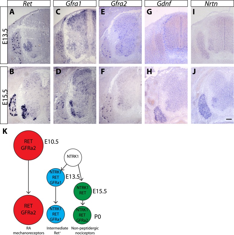
Figure 1—figure supplement 1. Expression of Ret, Gfras, and Gfls in developing spinal cord (SC) and DRG.

Figure 1—figure supplement 1.

(AJ) In situ hybridization of mouse SC and DRG at E13.5 and E15.5 for Ret (AB), Gfra1 (CD), Gfra2 (EF), Gdnf (GH), and Nrtn (IJ). Ret is expressed in DRG neurons and motor neurons at E13.5 and E15.5. Ret is also expressed in dSC from E15.5. Gfra1 is expressed in DRG neurons, motor neurons, dorsal root entry zone, and dSC at both stages. Note that expression of Gfra1 in the dorsal root entry zone and dSC is largely Ret independent. Gfra2 is expressed in large diameter DRG neurons and in motor neurons. Nrtn and Gdnf are barely detected in DRG and SC at E13.5 and display increased expression in DRGs at E15.5. (K) Schematic of temporal expression of Ret and Gfra co-receptors in DRG neurons, which is adapted from previous studies (Luo et al., 2007, 2009; Molliver et al., 1997). RA mechanoreceptors (red cells) are early RET+ DRG neurons, which begin to express Ret and Gfra2 from E10.5 or earlier. All other RET+ DRG neurons develop from NTRK1+ precursors and depend on NTRK1 signaling for their expression of Ret and Gfras. Intermediate RET+ neurons (blue cells) express Ret and Gfra1 from E13.5. The late RET+ non-peptidergic nociceptors express Ret from E15.5, and begin to express a low level of Gfra2 around P0. Scale bar = 100 μm.
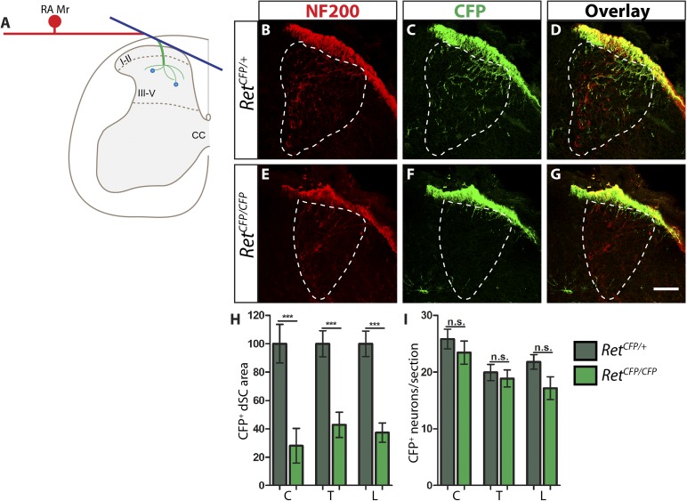
Figure 1—figure supplement 2. Ret is required for the growth of RA mechanosensory third order central projections at E13.5.

Figure 1—figure supplement 2.

(A) Schematic of development of RA mechanosensory central projections. RA mechanoreceptors grow central and peripheral axons soon after neurogenesis, generating first order branches (red color). Upon reaching the dSC, the central axons bifurcate and send second order longitudinal branches rostrally and caudally (blue color). Around E13.5, third order interstitial projections (green color) from the longitudinal branches innervate layers III–V of the dSC and develop complex branching patterns. Synaptic connections between mechanoreceptors and dSC neurons (light blue dots) develop from E18.5. (BG) Anti-NF200 and anti-GFP immunostaining of E13.5 RetCFP/+ (BD) and RetCFP/CFP (EG) SC. The dSC innervations of RA mechanosensory fibers are outlined by white dotted line. (H) Quantification of CFP+ pixel number in the dSC, which is displayed as a percentage of pixel number relative to control. There is a significant decrease in CFP+ axons innervating the SC in Ret mutants, suggesting that the initial growth of RA mechanosensory third order projections depends on RET signaling. (I) Quantification of the number of CFP+ neurons per DRG section. There is no significant change in the number of CFP+ neurons per DRG section, suggesting that there is no cell death or downregualtion of CFP expression in E13.5 Ret null RA mechanoreceptors. Scale bar = 50 μm. C = Cervical, T = Thoracic, L = Lumbar. Error bars represent SEM. n.s. = p > 0.05, *** = p < 0.001. Source data are provided in Figure 1—source data 2.
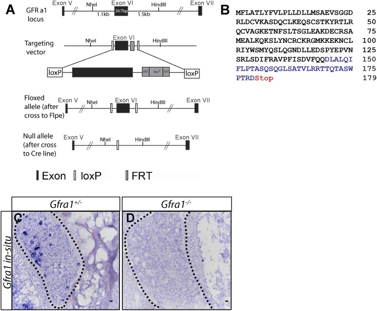
Figure 1—figure supplement 3. Generation of Gfra1 conditional and null alleles.

Figure 1—figure supplement 3.

(A) Schematic of generation of Gfra1 conditional and null alleles. See supplemental ‘Materials and methods’ for additional details. (B) Predicted peptide sequence of the truncated GFRa1 protein after the excision of exon 6. Black letters represent amino acids which share identity with wild-type protein sequence. The loss of exon 6 causes a frame shift, leading to the inclusion of amino acids which do not match the wild-type sequence (blue letters). The frame shift also introduces a premature stop codon following amino acid 179. (CD) In situ hybridization of Gfra1 in P0 Gfra1+/+ control (C) and Gfra1−/− null (D) DRG sections shows a complete loss of Gfra1 transcript in Gfra1 null tissue.