
Figure 5.
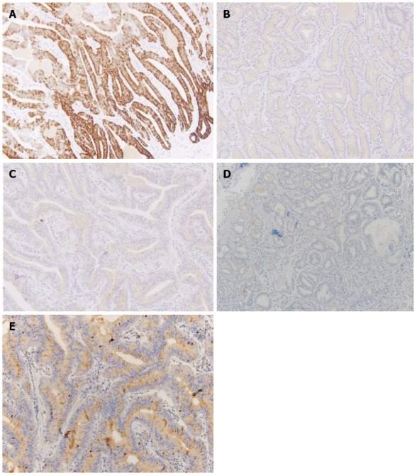
Immunohistochemical staining for mucin, α-fetoprotein, and Ki-67. A: Most of the glandular cells were positive for mucin (MUC) 6; B: Most of the glandular cells were negative for MUC2; C: Most of the glandular cells were negative for MUC5AC; D: Most of the glandular cells were negative for alpha fetal protein; E: Ki-67 immunolabeling demonstrates a low proliferation index (< 1%).