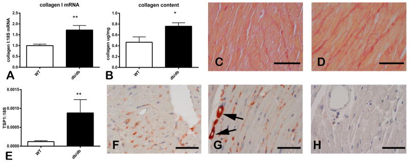
Figure 1. Cardiac fibrosis in the db/db myocardium is associated with TSP-1 upregulation.
A–D: db/db mice had significant fibrotic remodeling of the myocardium. qPCR analysis showed marked upregulation of collagen I mRNA in db/db hearts at 2 months of age (A). A hydroxyproline biochemical assay showed that 6-month old db/db mice had significantly higher myocardial collagen content when compared with age-matched WT animals (B). Sirius red staining shows increased deposition of collagen in the cardiac interstitium of db/db animals (D), when compared with lean WT mice (C). E: qPCR shows that TSP-1 mRNA expression was markedly increased in db/db hearts. F–G: Immunohistochemical staining of 6 month-old mouse hearts showed weak immunoreactivity in lean WT mice (F). In contrast, in the db/db myocardium, intense TSP-1 staining was noted (G) and was predominantly localized in perivascular and interstitial areas (arrows). TSP-1 KO mice showed negligible staining for TSP-1 (H) (*p<0.05 vs. WT; **p<0.01 vs. WT). Scale bar: 50μm.

