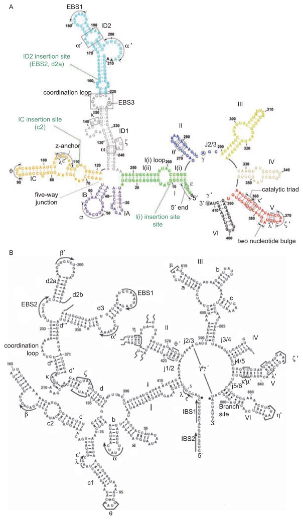
Figure 1.
Secondary structures of smaller (Group IIC) and larger (Group IIB) intron variants. (A) Secondary structure of the crystallized group IIC intron construct from Oceanobacillus iheyensis (O.i.). The sequence of the full-length O.i. intron is provided in Figure S2 of Toor et al. (2008). Domains are indicated with roman numerals and long-range interactions are indicated by Greek letters. Color coding is the same as shown in Figure 6, for the three-dimensional structure. Probable sites of the ID2, IC and I(i) insertions are indicated with green dotted lines, as indicated. (B) Secondary structure of the ai5γ Group IIB intron from the mitochondrial genome of Saccharomyces cerevisiae. Domains are indicated with roman numerals and subdomains with lower case letters. Long-range tertiary interaction partners are indicated with Greek letters. Note that EBS1 and EBS2 form Watson–Crick base pairs with IBS1 and IBS2 of the 5′-exon, respectively. Exon/intron boundaries are indicated by black dots.

