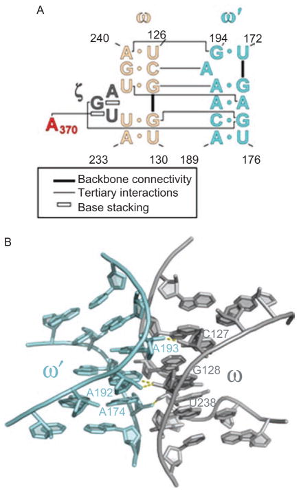
Figure 10.
The ω–ω′ interaction of group IIC introns. (A) Secondary structural representation of this sequence-specific ribose zipper motif. Color coding is as shown in Figures 1A and 6. First published in J Mol Biol (2008) 283, 475–481; Figure 1b, page 476. (B) Molecular structure of the ω–ω′ region within the Oceanobacillus iheyensis group IIC intron, showing the ribose zipper hydrogen bonds in yellow. First published in Science (2008) 320, 77–82; Figure 3c, page 79.

