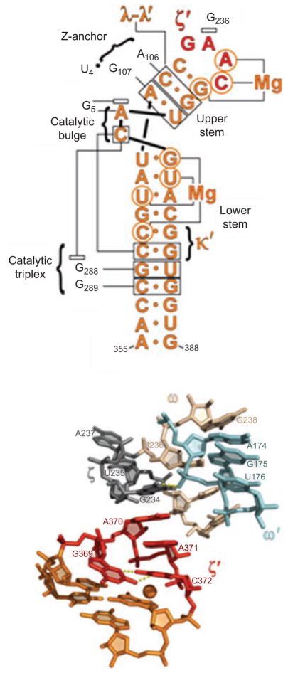
Figure 7.
The network of interactions involving DV. (A) A secondary structural map of the interactions observed between DV and the rest of the intron. Brackets indicate sites of long-range interaction and contacts with metals, open rectangles indicate stacked nucleobases, labels indicate identity of tertiary interactions. Red nucleotides are involved in the ζ–ζ′ interaction. (B) The molecular structure of the ζ–ζ′ interaction, determined from the crystal structure of the Oceanobacillus iheyensis group IIC intron. Unlike most tetraloop receptors, this interaction is dictated only by a single stack between A370 of DV and G236 in DI. The unusual tetraloop contains a bound metal ion. The receptor is buttressed by the ω–ω′ interaction that is idiosynchratic to IIC introns (Keating et al., 2008). First published in J Mol Biol (2008) 283, 475–481; Figures 1a and 2a, pages 476 and 478 respectively.

