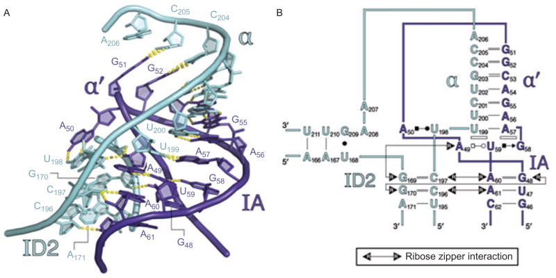
Figure 9.
The α–α′ interaction and its supporting ribose zipper network. (A) Molecular structure of α–α′, showing that the strands beneath it are joined through sequential contacts between 2′-hydroxyl groups (ribose zippers). (B) Secondary structural representation of the same region. Open rectangles indicate base stacking interactions. Types of base-pairing are represented using the Leontis–Westhof symbolism. Color coding is as shown in Figure 1A and 6. Reprinted with permission from RNA (Toor et al., 2010).

