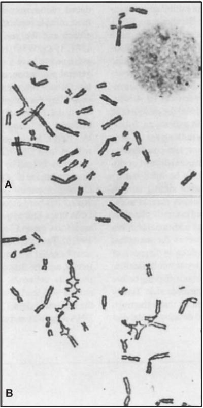
Figure 8.7.2.

Partial metaphase spreads from Fanconi anemia lymphocytes. (A) Untreated cell showing baseline chromatid aberrations. (B) Cell exposed to 0.1 μg/ml diepoxybutane (DEB). The multiple chromatid exchange figures seen at this concentration are unique to FA cells. Reproduced from Auerbach et al. (1981) with permission of the American Academy of Pediatrics.
