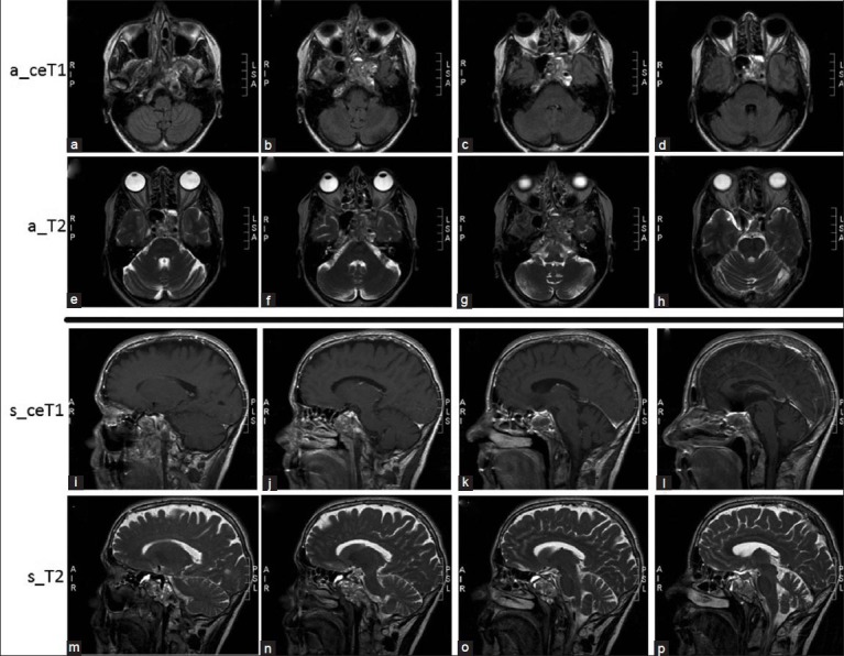
Figure 2.

Axial (a_) and sagittal (s_) contrast enhanced T1-weigthed (ceT1) and T2-weighted MRI images showing a heterogeneous lesion involving the clivus, (a, b, j, k, n, o) sphenoidal bone, (c, d, g, h, k, o) sphenoidal wing, (b, i) as well as extending to the left petrosal apex and cavernous (b, d, g, h, i, m) sinus
