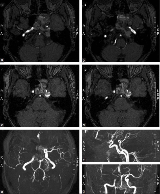
Figure 3.

MRI angiographic scans show the relationship between the tumor and major vessels of the skull base. The intracavernous and supraclinoidal segment of the left internal carotid was surrounded and compressed by the tumor (c, d, e, g). However, sufficient blood supply to the left medial and anterior brain arteries provided by the right carotid circulation through the Willis circle (e, f, g), as well as a slow tumor growth enabling collateralization, can explain the lack of neurological deficits besides the ones produced by a direct tumor compression of cranial nerves
