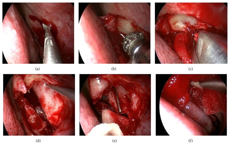
Figure 1.
EE-DCR. A square nasal mucosal flap was incised by a blade (a) and a power blur then was used to thin maxilla and frontal process of the maxilla (b), a rongeur to remove the bone (c), and a probe to bulge medial sac and allow the medial wall of the sac fully incised (d); finally the entire sac was opened (e) and the wound was packed with merogel (f).

