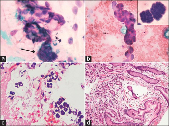
Figure 2.

A panel of microphotographs; (a) a cluster of atypical cells along with organisms present in tetrads and octads conforming to the morphology of Sarcina (H and E, ×100); (b) A small cluster of atypical epithelial cells; inset — Sarcina in tetrads and octads (H and E, ×100); (c) biopsy showing the organism Sarcina (H and E, ×40); (d) biopsy showing the presence of tumor cells in the lamina propria (H and E, ×40)
