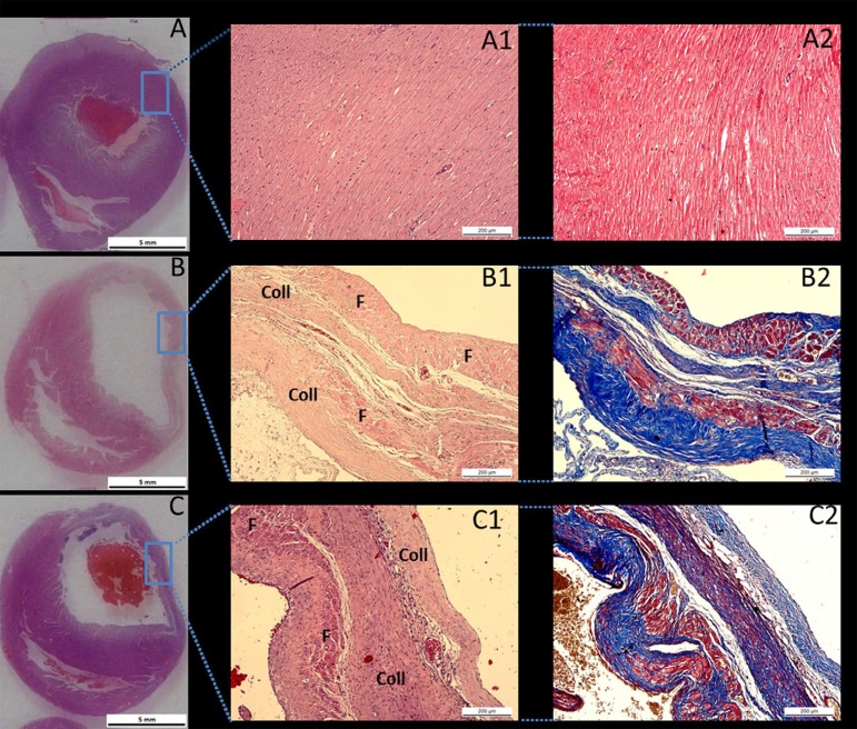
Fig. 3.
Panoramic photomicrographs of the cross-sections of the heart from a SHAM rat (A), a G1 rat (B), and a G2 rat (C).
*In the G1 and G2 rats, a transmural infarct is observed on the free wall of the left ventricle. Haematoxylin and eosin staining of cardiac tissue from a SHAM rat (A1) in which no myocardial infarct was observed, the myocardium is preserved and no histological alterations in cardiac microarchitecture were found. Haematoxylin and eosin staining of cardiac tissue in a G1 rat (B1) and a G2 rat (C1) shows an organized area of cicatricial collagen (Coll) in the transmural infarct of remanescent muscle fibres (F). Masson trichrome staining of cardiac tissue from a SHAM rat (A2), a G1 rat (B2), and a G2 rat (C2) in which the muscle fibres are stained red and the collagen is stained blue

