Figure 1.
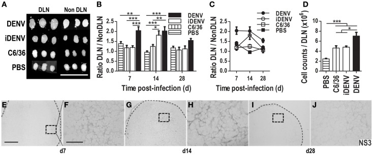
Cutaneous inoculation of DENV infects immune-competent mice inducing an immune response. The size of draining lymph nodes (DLN) and non-DLN (contralateral inguinal LN) was compared among groups of immune-competent BALB/c mice inoculated with either intact DENV, UV-inactivated-DENV (iDENV), supernatant from uninfected C6/36 mosquito cell line (C6/36), or endotoxin-free PBS. (A) DENV induces the biggest DLNs post-inoculation (14 days post-inoculation). (B,C) The ratio between the sizes of these LN (DLN/Non -DLN) at days 7, 14, and 28 post-inoculation. (D) The cellularity obtained per DLN at day 14 post-inoculation was higher in DENV inoculated mice than in mice inoculated with the other conditions. Data shown are pooled from three independent experiments, and represent the mean ± SEM. (E–J) NS3 viral protein was identified by means of immunohistochemistry (IHC, DAB, gray color) in DLN from DENV-infected mice at different time points post-inoculation [day 7 (E,F); day 14 (G,H); day 28 (I,J)]. Pictures in (F,H,J) (60×, scale bar 40 μm) correspond to the squares indicated on the left: (E,G,I) (10×, scale bar 200 μm). Representative pictures from one of three independent experiments are shown. Mice were inoculated i.d. with 6 × 10⁴ pfu of DENV or the other conditions at day 0, boosted at day 7, and sacrificed at the indicated days. Dotted lines depict the limits of LNs. Data were analyzed with unpaired two-tailed Student’s t-test, and two-way ANOVA with the Bonferroni post-test. *P < 0.05, **P < 0.01, ***P < 0.001.

