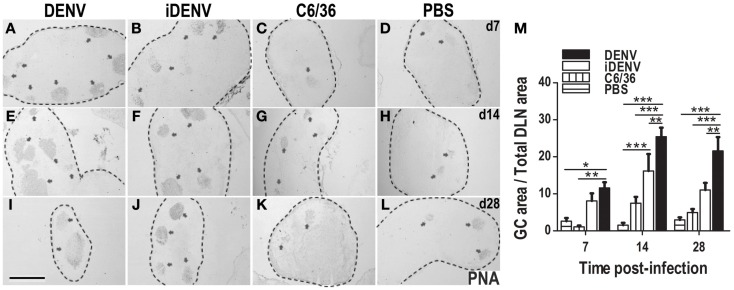
Figure 2.
Cutaneous DENV induces an increased number and size of GCs compared with control conditions. (A–L) DLNs from mice inoculated i.d. with DENV (A,E,I), iDENV (B,F,J), C6/36 (C,G,K), or PBS (D,H,L) and collected at 7 (A–D), 14 (E–H), and 28 (I–L) days post-inoculation show an increased number and bigger GCs in animals infected with DENV compared with the other conditions. The limits of LN are indicated by dotted lines. Arrowheads indicate GCs, labeled in situ with PNA and revealed in blue-gray color (SG Vector). Representative pictures from one of three independent experiments are shown. Mice were inoculated at day 0 and boosted at day 7. (M) The kinetic of GCs area as percentage of GCs Area over Total LN Area in each condition is shown. Data shown are pooled from three independent experiments, represent the mean ± SEM, and were analyzed with two-way ANOVA with the Bonferroni post-test. *P < 0.05, **P < 0.01, ***P < 0.001. Scale bar, 500 μm.

