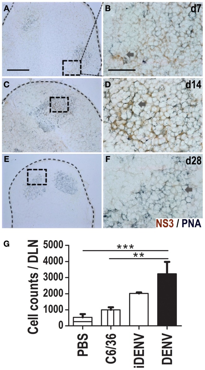
Figure 6.
NS3 viral protein is present inside GCs in DLNs. Viral NS3 protein was identified in situ (DAB, brown color) in a double staining with GCs (PNA, blue-gray color) in DLN at day 7 (A,B), 14 (C,D), and 28 (E,F) post-DENV-inoculation. Positive cells were found inside GCs. For each time point, right images [60×, scale bar 40 μm; (B,D,F)] are derived from left indicated squares [10×, scale bar 200 μm; (A,C,E)]. Data are representative from three independent experiments. Dotted lines indicate the limits of LNs. (G) NS3-specific GC B cells at day 14 post-inoculation by flow cytometry using multimers of NS3 viral protein. Data shown are pooled from two independent experiments, represent the mean ± SEM, and were analyzed with two-way ANOVA with the Bonferroni post-test. **P < 0.01, ***P < 0.001. Mice were inoculated at day 0 and boosted at day 7.

