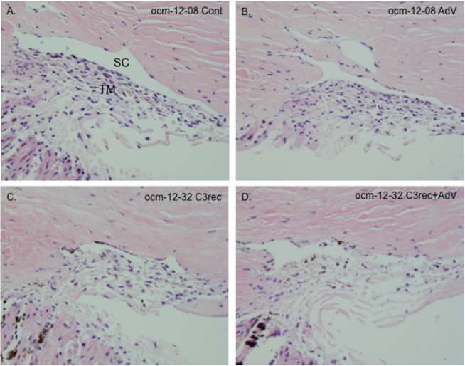
Figure 4.
H&E staining showing gross morphology of segments at 24 hours after the various treatment regimens. (A, B) Untreated control (Cont) and AdV treated segments from MOCAS ocm-12-08. There were no apparent qualitative morphological differences between paired control and AdV-treated segments in terms of cellularity of the juxtacanalicular regions and along the collagen beams, the integrity of Schlemm's canal (SC), and the organization of the collagen beams. (C, D) The C3rec- and C3rec + AdV–treated segments from MOCAS ocm-12-32. There was reduced cellularity, disorganized beams, and discontinuity of the inner wall of SC. These alterations appeared to be more prominent in the AdV + C3rec segment than in the C3rec segment. TM, trabecular meshwork.

