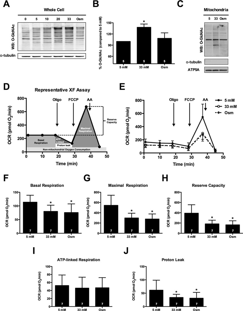
Figure 1. High glucose depresses the bioenergetic reserve capacity of cardiomyocytes.
(A) Immunoblot of whole-cell protein O-GlcNAcylation following 0, 5, 10, 20 and 33 mM glucose treatment. An osmotic control (Osm, 5 mM glucose + 28 mM mannitol) was also used. (B) Densitometric measurement of O-GlcNAcylation in relation to 5 mM control demonstrated a significant induction of protein O-GlcNAcylation following 33 mM glucose. (C) Immunoblotting for mitochondrial protein O-GlcNAcylation. NRCMs were fractionated to isolate mitochondrial protein following treatment with 5 mM glucose, 33 mM glucose and the osmotic control. (D) Representative XF assay and diagram of how mitochondrial measurements were calculated. (E) Mitochondrial function assay following 48 h of high glucose treatment. From (E) parameters of mitochondrial function were assessed (as described in D): (F) basal respiration, (G) maximal respiration, (H) reserve capacity, (I) ATP-linked respiration, and (J) proton leak. As indicated in bars, n = 7 independent experiments; *P < 0.05, compared with 5 mM.

