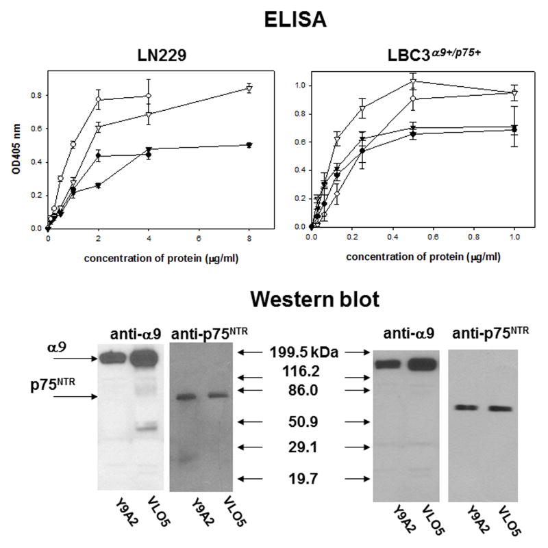
Fig. 2.
Detection of the α9/p75NTR complex in preparations obtained from affinity chromatography. Lysates of LN229 and LBC3α9+/p75+ cells were applied to affinity columns containing resins coupled with VLO5 or Y9A2. The retained proteins were eluted with 5 mM EDTA from the VLO5 column and low pH = 2.7 from the Y9A2 mab column. Identification of α9βl integrin and p75NTR was performed in ELISA with different concentrations of immobilized proteins obtained from the VLO5 column (circles) and Y9A2 column (triangles). Y9A2 mab was used for detection of the integrin (open symbols) and ME20.4 mab for detection of p75NTR (filled symbols). Error bars represent the standard deviation from three independent experiments. Western blot analysis of the purified proteins was performed using anti-α9 polyclonal antibody and anti-p75NTR mab.

