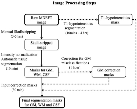
Figure 1.

This flow chart represents the image processing steps that were followed to obtain final segmentation for masks gray matter (GM), white matter (WM), and CSF. Raw MDEFT images were manually deskulled, intensity normalized, and automatically segmented into GM, WM, and CSF. The obtained segmentation masks were corrected for misclassifications of deep GM and T1 hypointensities. The generated GM correction mask and T1 hypointensities mask were used to correct the original tissue masks, thus obtaining final corrected segmentations for GM, WM, and CSF.
