Fig 3.

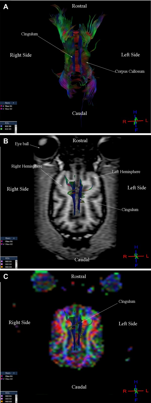
Images illustrating the cingulum in a healthy dog. (A) Diffusion tensor tractography (DTT) image, dorsal view. (B) T1-weighted image and DTT image, dorsal view. (C) Diffusion tensor tractography image on the colored map, dorsal view.

Images illustrating the cingulum in a healthy dog. (A) Diffusion tensor tractography (DTT) image, dorsal view. (B) T1-weighted image and DTT image, dorsal view. (C) Diffusion tensor tractography image on the colored map, dorsal view.