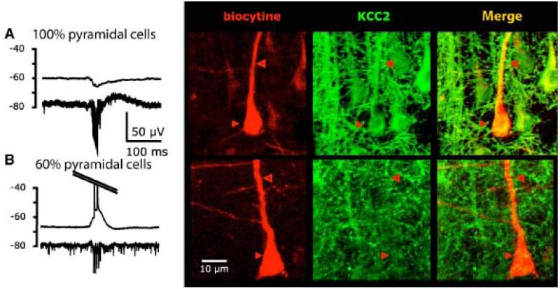
Figure 5.
Correlation between the behavior of a pyramidal cell during interictal discharges (hyperpolarization in a A vs depolarization in B) and the expression of KCC2 (based on Huberfeld et al., 2007). Electrophysiological trace is shown on the left (top: intracellular recording – bottom: extracellular). The recorded cell is then filled with biocytin and KCC2 is labeled by immunohistochemistry.

