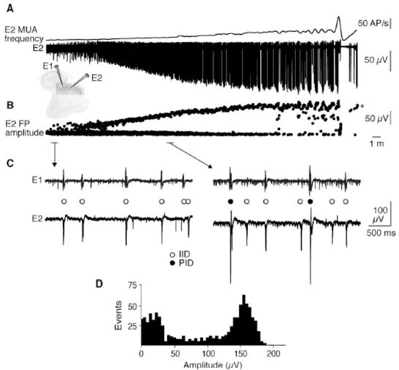
Figure 8.
Preictal discharges progressively emerge during the transition to the seizure.
A: Extracellular recordings of the transition to seizure-like activity. E1 and E2 are recordings from two subicular electrodes. MUA frequency (upper trace) and the extracellular signal from E2 (lower trace) are shown.
B: Amplitude measurements for all field potentials recorded by electrode E2 during the transition show the emergence of larger PIDs, whereas the amplitude of inter-ictal events did not change.
C: Dual extracellular recordings showing IIDs (open circles, left) before convulsant application and coexpression of PIDs (filled circles, right) with IIDs during the transition.
D: Amplitude distribution for all field potentials during the 35-min transition period showed IIDs of amplitude 10–50 μV and PIDs of amplitude 125–175 μV.

