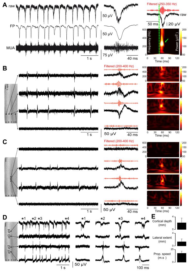
Figure 2. Interictal-like discharges are spatially restricted to superficial neocortical layers and are generated in multiple sites.
(A) Extracellular records of interictal-like discharges (IIDs). They consist of bursts of multi-unit activity (MUA; > 300 Hz high pass filter) correlated with field potentials (FP, < 100 Hz low pass filter) (left). Expanded raw, MUA and FP traces at middle (middle). High frequency oscillations (HFOs; filtered to pass 250–350 Hz) are detected during IIDs (right). Time frequency plots (below) show a mean dominant frequency of 251±69 Hz (n=572 events).
(B, C) Multiple extracellular records of IIDs from slices containing infiltrated neocortex. In B, four electrodes are placed at different depths of the same neocortical column. They show IIDs and HFOs were synchronous in a vertical column-like region with field onset, maximal amplitude and the largest HFO in layers III-IV. In C, four electrodes are placed in layers IIIIV at the same depth over a lateral distance of 1 mm. They show a spatially restricted lateral spread of IIDs and HFOs. HFOs were never detected in the absence of IIDs.
(D) IIDs are initiated at multiple foci in infiltrated neocortex. Multiple records from a slice of infiltrated neocortex with four electrodes (el) located in layers III-IV at separations of: el1 – 1mm – el2 – 3mm – el3 – 1mm – el4. Expanded traces in the middle.
(E) Mean±SD of the neocortical depth, lateral extent and propagation speed of IIDs in the recorded slices.

