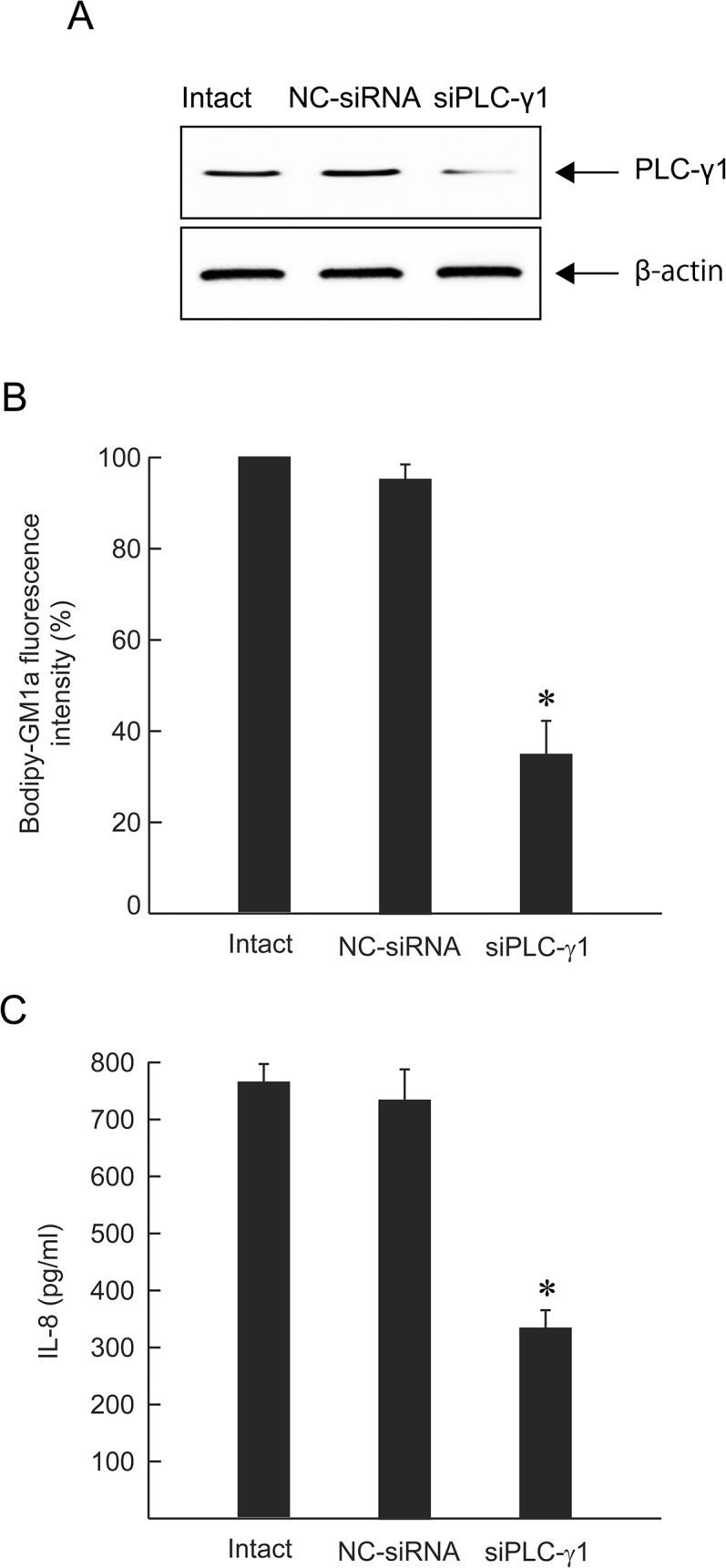
Fig 7. Effect of siRNA on clustering of GM1a and release of IL-8.

A549 cells were transfected with siPLCγ-1 or NC-siRNA (10 nM). (A) Expression of PLCγ-1 was detected by western blotting with anti-PLCγ-1 and anti-β-actin antibodies. (B) Intact cells, NC-siRNA-treated cells, or siPLCγ-1-treated cells were stained with BODIPY-GM1a and incubated with 1.0 μg/mL alpha-toxin at 37°C for 60 min. The cells were fixed in 4% paraformaldehyde and analyzed by fluorescence microscopy. Fluorescence intensity was measured. The clustering of GM1a in the intact cells was set as the maximal response (100%) against which all other results were compared. Values represent the mean ± SE; n = 3; *, p < 0.01. (C) siRNA-treated cells were incubated with 1.0 μg/mL alpha-toxin at 37°C for 3 h. The concentration of IL-8 in the culture supernatants was determined by ELISA. Values represent the mean ± SE; n = 5; *, p < 0.01.
