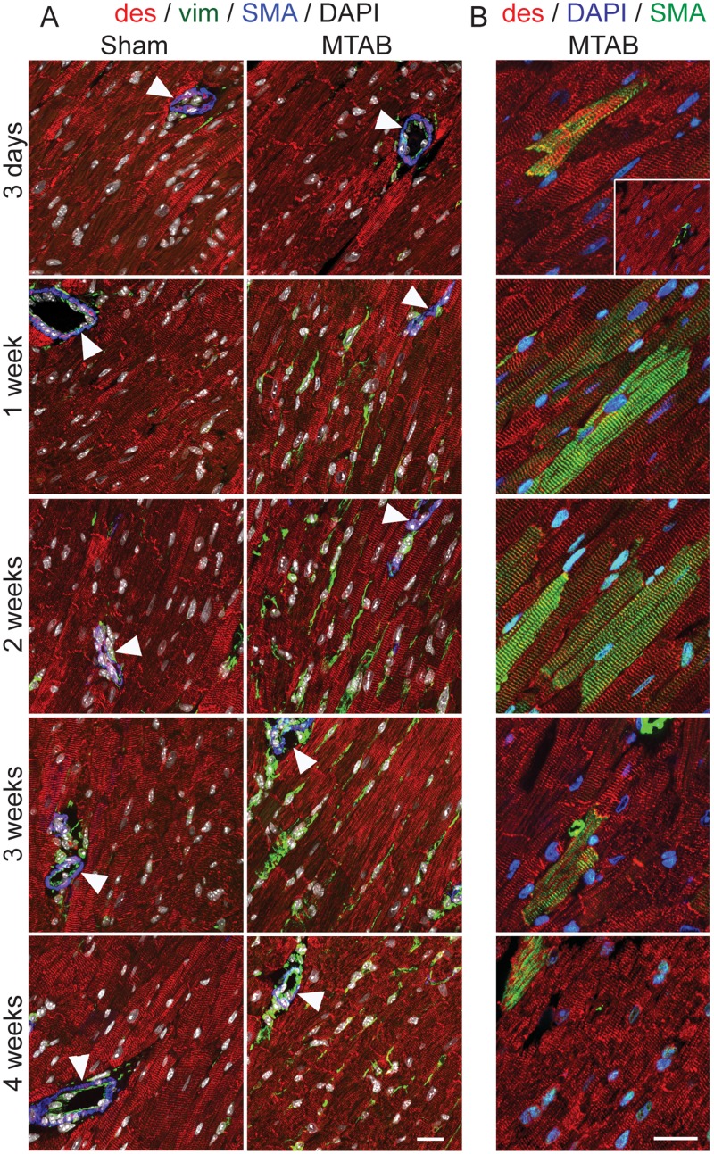
Fig 3. Cellular responses to PO in the myocardium.
WT mice were Sham operated or subject to aortic banding (MTAB) and sacrificed after the indicated time points. Transmural blocks of the left ventricular myocardium were sectioned and immunostained for (A) α-SMA (SMA, blue), desmin (des, red), vimentin (vim, green) and nuclei (DAOI, white) or (B) α-SMA (SMA, green), desmin (des, red), and nuclei (DAOI, blue). Note that exposure time for α-SMA imaging was adjusted in (A) to the high staining intensity in vascular smooth muscle and in (B) to the comparably lower staining intensity in cardiomyocytes overloaded myocardium (~2-times longer exposure). Scale bars: 50 μm.

