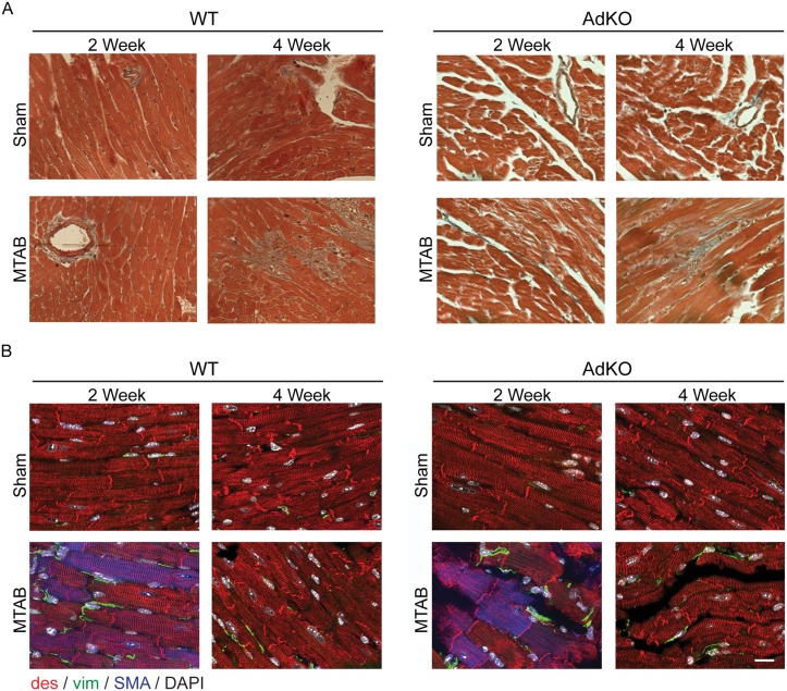
Fig 4. Effects of MTAB in WT and AdKO animals on myocardial cells.
(A) Masson’s trichrome staining of histological sections taken from Ad KO or WT mice after 2 or 4 weeks following sham or MTAB surgery. Images shown are representative of 5–10 images of n = 4 to 6 mice per group. (B) WT and AdKO (KO) mice were Sham operated or subject to aortic banding (MTAB) and sacrificed after 2 and 4 weeks post PO. Transmural blocks of the left ventricular myocardium were sectioned and immunostained for α-SMA (SMA, blue), desmin (des, red), vimentin (vim, green) and nuclei (DAPI, white). Scale bar: 50 μm.

