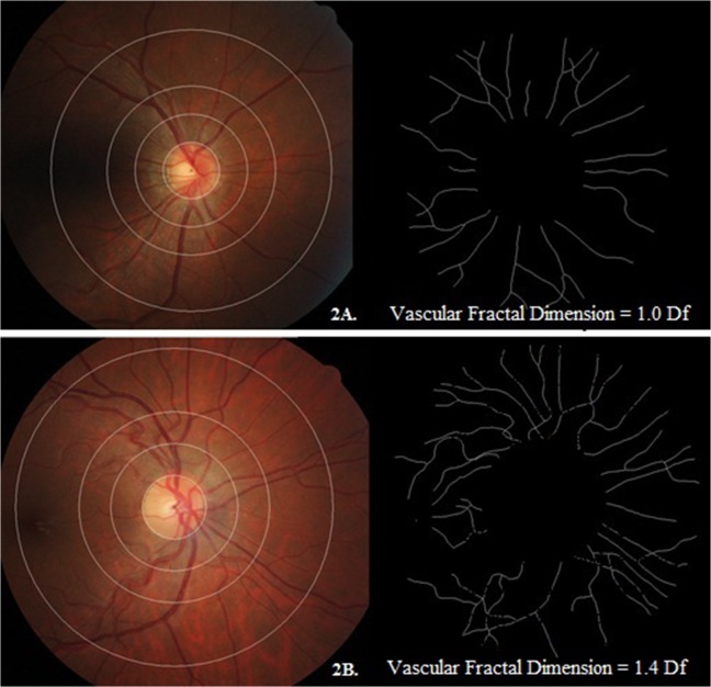
Fig 2. Examples of retinal fundus photographs with relevant pattern of retinal vascular fractal dimension in our cohort.
The fractal dimension was shown in black and white images. The retinal vascular fractal dimension of Fig 2A and 2B were 1.0 Df and 1.4 Df, respectively. Compared with mother of Fig 2B, mother of Fig 2A showed a sparser retinal vascular fractal dimension.

